
What is glaucoma, and how do I know if I have it?
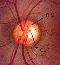
A normal optic nerve has uniform, pink, healthy rim tissue and a small cup.
Weinreb, R.N.
Glaucoma is a disease that causes damage to the optic nerve, which is in the back of the eye. The optic nerve carries visual information to the brain. Glaucomatous damage to this nerve results in vision loss that starts in the peripheral vision and progresses toward the central vision. In most cases this damage occurs due to fluid build up inside the eye which results in increased eye pressure. The exact cause of this fluid build up is unknown, and it cannot be prevented.
Glaucoma is a leading cause of blindness in the U.S., but if caught early it can be treated and further vision loss can be prevented. Unfortunately, glaucoma has no symptoms; however it can be detected with regular optometric examinations. Anyone can develop glaucoma, but it is more common in people who are over age 40, who are of African decent, who have a family history of glaucoma or who are very nearsighted.
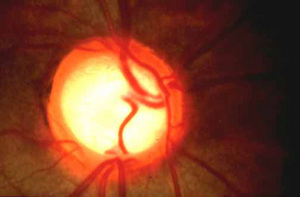
An optic nerve with glaucoma has thin rim tissue and a large cup.
Eyedoctor.homestead.com
There are four major types of glaucoma: open angle glaucoma, acute angle closure glaucoma, congenital glaucoma and secondary glaucoma. Open angle glaucoma is the most common form of the disease. In this form, the eye pressure increases slowly over time, damaging the optic nerve, causing gradual, painless vision loss. This type of glaucoma tends to run in families and is more common in blacks than in whites. It has no symptoms in early stages.
Acute angle closure glaucoma occurs when fluid outflow is suddenly blocked, causing a quick, severe, painful increase in eye pressure. This type of glaucoma is an emergency, because it can cause permanent vision loss very quickly. It is usually accompanied by symptoms of blurred vision, loss of peripheral vision, appearance of colored halos around lights, pain or redness in the eye and nausea. If acute angle closure develops in one eye, the other eye is at risk for developing it, so preventative treatment is recommended in the fellow eye.
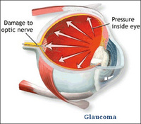
Increased eye pressure causes damage to the optic nerve.
Stuart MacFarlane
Congenital glaucoma is hereditary and is present at birth. It results from improper development of the fluid outflow channels in the eye. Secondary glaucoma can be caused by medications such as corticosteroids, eye conditions such as uveitis or systemic conditions.
Since most forms of glaucoma produce no symptoms until the disease is advanced and vision loss is severe, regular eye examinations are an important means of early detection. Eye exams should include tonometry, ophthalmoscopy and visual field testing. Tonometry is a measure of the pressure inside the eye. Glaucoma cannot be diagnosed on eye pressure alone because it changes.

Glaucoma causes loss of peripheral vision. This is how someone with advanced glaucoma might see.
Center for Sight
Also, one form of glaucoma occurs at normal pressures; it is called normal-tension glaucoma. Ophthalmoscopy is an examination of the back of the eye and an assessment of optic nerve health. A visual field test checks for abnormal blind spots in the vision.
There is no cure for glaucoma, but, in most cases, it can be successfully treated with eye drops that lower the eye pressure. Sometimes surgery may be necessary. Any vision loss that occurs due to glaucoma cannot be restored. But early detection, prompt treatment and careful monitoring by an optometrist can help most glaucoma patients maintain their normal daily lifestyle.
Dr. Jamie Barnes is an associate optometrist at Bennett Optometry. Bennett Optometry has two offices in Ann Arbor. Please visit our Web site at www.bennettoptometry.com.